
Mouse Secondary Antibodies


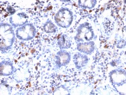
Biotium MUC3 (Mucin 3)(MUC3/2992R), CF568 conjugate, 0.1mg/mL
Recognizes a protein of HMW, identified as mucin 3 glycoprotein (MUC3). Its epitope localizes between aa SITTTE. This MAb shows no cross-reaction with human milk fat globule membranes, MUC1, or MUC2. MUC3 is distributed in colon and rectum, and is also present to a lesser extent in breast, lung and salivary gland tissues. The Mucins are a family of highly glycosylated, secreted proteins with a basic structure consisting of a variable number of tandem repeats (VNTRs) encoded by 60 base pairs (Mucin 1), 69 base pairs (Mucin 2) and 51 base pairs (Mucin 3). The number of repeats is highly polymorphic and varies among different alleles. Mucin 1 proteins are expressed as type I membrane proteins in addition to secreted forms. Mucin 1 is aberrantly expressed in epithelial tumors including breast carcinomas. Mucin 2 coats the epithelia of the intestines and airways and is associated with colonic tumors. Mucin 3 is a major component of various mucus gels and is broadly expressed in no

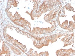
Biotium CD47 / IAP (Integrin Associated Protein)(CD47/3019), CF740 conjugate, 0.1mg/mL
This antibody reacts with Ig domain of CD47 protein. CD47, originally named integrin-associated protein (IAP), is a 50 kDa protein containing five membrane-spanning sequences and a short cytoplasmic tail. CD47 plays a role in both cell adhesion by acting as an adhesion receptor for THBS1 on platelets, and in the modulation of integrins. It is important in memory formation and synaptic plasticity in the hippocampus. CD47 may play a role in membrane transport and/or integrin dependent signal transduction. Primary antibodies are available purified, or with a selection of fluorescent CF Dyes and other labels. CF Dyes offer exceptional brightness and photostability. Note: Conjugates of blue fluorescent dyes like CF405S and CF405M are not recommended for detecting low abundance targets, because blue dyes have lower fluorescence and can give higher non-specific background than other dye colors.
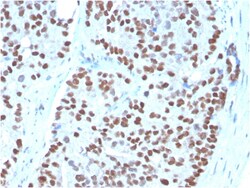
Biotium NKX3.1 (Metastatic Prostate Adenocarcinoma Marker)(NKX3.1/3348), CF568 conjugate, 0.1mg/mL
NKX3. 1 is a prostate specific gene encoding a transcription factor that plays an important role in normal prostate development and carcinogenesis. It is a prostatic tumor suppressor gene located on chromosome 8p21. 2, which frequently undergoes a loss of heterozygosity. NKX3. 1 expression is highly restricted in prostate epithelial cells and therefore can be used as a diagnostic biomarker for prostate cancer and other metastatic lesions of prostatic origin. Furthermore, NKX3. 1 shows better sensitivity than Prostate Specific Antigen (PSA) for identifying metastatic prostatic adenocarcinoma. This suggests that immunohistochemical staining of NKX3. 1, along with other prostate-restricted markers, may be valuable for the definitive determination of prostatic origin in poorly differentiated metastatic carcinomas. Primary antibodies are available purified, or with a selection of fluorescent CF Dyes and other labels. CF Dyes offer exceptional brightness and photostability. Note
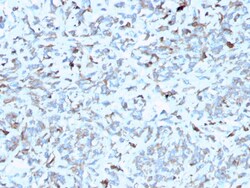
Biotium C1QA / Complement C1q A-Chain(C1QA/2952), CF568 conjugate, 0.1mg/mL
Complement C1q A Chain (C1qa) is a subunit of C1q. C1q is composed of 6 polypeptide chains each of C1qa, C1qb, and C1qc. C1q is a component of the classical complement pathway, and thereby plays an important role in the immune response. Each C1q subunit contains an N-terminal collagen-like region and a C-terminal C1q globular domain. The presence of receptors for C1q on effector cells modulates its activity, which may be antibody-dependent or independent. Macrophages are the primary source of C1q, while anti-inflammatory drugs as well as cytokines differentially regulate expression of the mRNA as well as the protein. C1q deficiency is associated with lupus erythematosus and glomerulonephritis. Primary antibodies are available purified, or with a selection of fluorescent CF Dyes and other labels. CF Dyes offer exceptional brightness and photostability. Note: Conjugates of blue fluorescent dyes like CF405S and CF405M are not recommended for detecting low abundance targets,
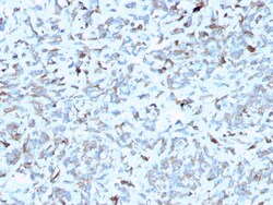
Biotium C1QA / Complement C1q A-Chain(C1QA/2952), CF740 conjugate, 0.1mg/mL
Complement C1q A Chain (C1qa) is a subunit of C1q. C1q is composed of 6 polypeptide chains each of C1qa, C1qb, and C1qc. C1q is a component of the classical complement pathway, and thereby plays an important role in the immune response. Each C1q subunit contains an N-terminal collagen-like region and a C-terminal C1q globular domain. The presence of receptors for C1q on effector cells modulates its activity, which may be antibody-dependent or independent. Macrophages are the primary source of C1q, while anti-inflammatory drugs as well as cytokines differentially regulate expression of the mRNA as well as the protein. C1q deficiency is associated with lupus erythematosus and glomerulonephritis. Primary antibodies are available purified, or with a selection of fluorescent CF Dyes and other labels. CF Dyes offer exceptional brightness and photostability. Note: Conjugates of blue fluorescent dyes like CF405S and CF405M are not recommended for detecting low abundance targets,
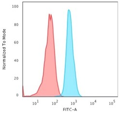
Biotium Cytokeratin 15 (Esophageal Squamous Cell Carcinoma Marker)(KRT15/2958), CF568 conjugate, 0.1mg/mL
Keratin 15 is a type I keratin which is expressed only in basal keratinocytes in stratified epithelia and does not appear to have a natural type II expression partner. Keratin 15 is down regulated in activated keratinocytes. Cytokeratin 15 is a specific marker of stem cells of the hair-follicle bulge and may be a useful marker for diagnosis between basal cell carcinoma (BCC) and trichoepithelioma. Trichoblastoma are benign neoplasms of follicular differentiation frequently found in nevus sebaceous. Many morphologic features are shared with nodular basal cell carcinoma, sometimes rendering a diagnosis difficult. Trichoblastoma and BCC show variable expression of Cytokeratin 15 and Cytokeratin 19, and absence of hair keratins. Primary antibodies are available purified, or with a selection of fluorescent CF Dyes and other labels. CF Dyes offer exceptional brightness and photostability. Note: Conjugates of blue fluorescent dyes like CF405S and CF405M are not recommended for
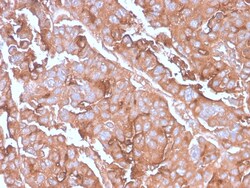
Biotium MUC1 / CA15-3 / EMA / CD227 (Epithelial Marker)(MUC1/2980R), CF740 conjugate, 0.1mg/mL
This MAb recognizes full-length MUC1 in a glycosylation-independent manner and can bind to the fully glycosylated protein. The dominant epitope of this MAb is APDTR in the VNTR region. It reacts with the core peptide of the MUC1 protein, which is a member of a family of mucin glycoproteins that are characterized by high carbohydrate content, O-linked oligosaccharides, high molecular weight (200 kDa) and an amino acid composition rich in serine, threonine, proline and glycine. The core protein contains a domain of 20 amino-acid tandem repeats that functions as multiple epitopes for the MAb. Incomplete glycosylation of some tumor-associated mucins may lead to variable unmasking of the multiple peptide epitopes leading to the observed differences in staining intensity between normal and malignant tissues. This MAb reacts with both normal and malignant epithelia of various tissues including breast and colon. Primary antibodies are available purified, or with a selection of fluore
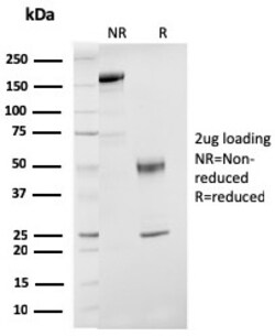
Biotium ER-beta-1 (Estrogen Receptor beta-1)(ESR2/3207), CF488A conjugate, 0.1mg/mL
Estrogen receptors (ER) are members of the steroid/thyroid hormone receptor superfamily of ligand-activated transcription factors. Estrogen receptors, including ER-alpha and ER-beta, contain DNA binding and ligand binding domains and are critically involved in regulating the normal function of reproductive tissues. They are located in the nucleus, though some estrogen receptors associate with the cell surface membrane and can be rapidly activated by exposure of cells to estrogen. ER-alpha and ER-beta are differentially activated by various ligands. Receptor-ligand interactions trigger a cascade of events, including dissociation from heat shock proteins, receptor dimerization, phosphorylation and the association of the hormone activated receptor with specific regulatory elements in target genes. Evidence suggests that ER-alpha and ER-beta may be regulated by distinct mechanisms even though they share many functional characteristics. Primary antibodies are available purified, o
Biotium CD11c (Dendritic Cell Marker) ( ITGAX/1243) , CF405S conjugate, 0.1mg/mL
This antibody recognizes a protein of 145 kDa, identified as CD11c. CD11c (ITGAX), a member of the leukointegrin family, shares the same beta subunit with other members of the leukocyte adhesion molecule family, which includes CD11a (LFA-1), CD11b (MAC-1) and CD11d (ITGAD), but has a unique alpha chain. CD11c has been shown to play a role in phagocytosis, cell migration, and cytokine production by monocytes/macrophages as well as induction of T-cell proliferation by Langerhans cells. CD11c is expressed prominently on the plasma membranes of monocytes, tissue macrophages, NK cells, and most dendritic cells (DCs). A lower level of expression is also observed on neutrophils as a result of its high level of expression on most DCs. An antibody to CD11c may aid in identification of lesions with histiocytic origin. It may also been used as a marker for hairy cell leukemia in paraffin-embedded tissues.Primary antibodies are available purified, or with a selection of fluorescent CF Dyes a